What is lobar pneumonia?
Lobar pneumonia is a lung infection that affects an entire lung lobe or a large and continuous part of it [1]. When it affects more than one lobe, it is called multilobar, and when it affects all lung lobes, it is called panlobar pneumonia.
Causes
Lobar pneumonia is most often caused by bacteria, such as Streptococcus pneumoniae or Haemophilus influenzae, but also by other bacteria, such as Klebsiella, Mycoplasma or Legionella, or viruses [1].
Histology
Lobar pneumonia has 4 stages [1]:
- Congestion in the first 24-48 hours with engorgement of the lung blood vessels and fluid in the air sacs (alveoli).
- Red hepatization or consolidation (2nd-4th day) with fluid, red cells, white cells called neutrophils, and fibrin in the air sacs. Under the microscope, the lung tissue has the appearance of liver tissue (in Latin, liver = hepar, so from here the term hepatization)
- Grey hepatization (4th-6th day) with breakdown of red liver cells and persistence of neutrophils and fibrin in the air sacs
- Resolution (8th-9th day) = complete recovery
Symptoms and Signs
Symptoms of lobar pneumonia usually develop suddenly and can include coughing up yellow, green or rusty mucus, shortness of breath, high fever, fatigue and chest pain during coughing.
During percussion (tapping the chest with a finger), a doctor can hear a dull sound and during listening the lungs (auscultation), crackling sounds and decreased breathing sounds over the affected lung lobe.
X-Ray
On an X-ray, lobar pneumonia appears as a continuous white patch in a lung lobe.
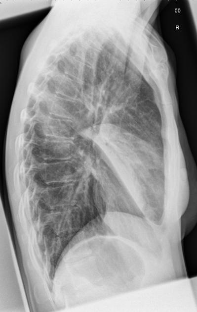
Picture 1. Lobar pneumonia in the middle lobe
of the right lung – an X-ray image from the right side,
showing a triangular white patch (source: Radiopedia, CC license)
NOTE: The pattern of lobar pneumonia on X-ray film does not already mean a bacterial cause.
Treatment
Treatment of lobar pneumonia depends on the cause — bacterial pneumonia is treated with antibiotics and viral pneumonia by antivirals.
Lobar Pneumonia vs Bronchopneumonia
Lobar pneumonia affects an entire lung lobe, usually only in one part of the lung; an X-ray usually shows a single solid white patch (or two or more, if more lobes are affected).
Bronchopneumonia affects small breathing ways (bronchioli) and small parts of the lung tissue around them (lobuli); an X-ray shows small white patches scattered over the large area of, usually both, lung wings.
- References
- Roy S, Pathology of Pneumococcal Pneumonia (Lobar Pneumonia) Histopathology-India.net