What is the gallbladder?
The gallbladder is a small hollow organ that stores the bile produced by the liver and delivers it into the duodenum.
The medical terms for the gallbladder include:
- Vesica biliaris or vesica fellea (from Latin vesica = bladder; bilis or fel = bile) [4,17]
- Cholecyst (from Greek khole = bile; kystis = bladder) [16]
Can you live without the gallbladder?
Yes, you can live without the gallbladder since it is not a vital organ. The gallbladder does not produce the bile; it only stores it. After gallbladder removal, the bile constantly flows from the liver into the intestine, which can cause diarrhea in some people.
Gallbladder agenesis is a rare condition in which a person is born without the gallbladder but usually has no symptoms [6].
Where is the gallbladder in your body?
The gallbladder lies in the upper right abdominal quadrant under the cartilage of the 9th rib and is covered by the lower edge of the liver [12]. It lies about an inch medially from an imaginary vertical line from the right breast nipple.

Picture 1. Gallbladder lies beneath the lower liver edge
at the bottom of the rib cage.
The intrahepatic gallbladder is an anatomical variant in which the gallbladder is completely embedded into the liver [10].
Anatomy
Shape and Size
Gallbladder measures in adults:
- Shape: pear-like
- Length: 3-4 inches (7-10 cm) [1,12]
- Width: 1-1.5 inch (2.5-3.8 cm) [1,12]
- Weight: ~2.5 ounces (~70 grams) [3]
- Volume: 30-50 mL [5,12]
Some people have two gallbladders with either single or two cystic ducts [5].
Parts
The gallbladder is a muscular sac, which can be divided into the fundus, body and neck (infundibulum).

Picture 2. Gallbladder parts and bile ducts
Bile Ducts
The cystic duct connects the gallbladder to the common hepatic duct; the ducts merge and form the common bile duct (Picture 2). All the ducts are made of the fibroelastic tissue, not muscular tissue [5].
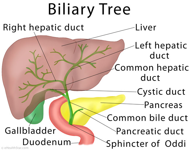
Picture 3. Biliary system: the gallbladder and bile ducts
Blood Supply
The flow of the arterial blood: aorta > celiac trunk > common hepatic artery > right proper hepatic artery > cystic artery [5].
The flow of the venous blood: cholecystohepatic veins > portal vein [5].
Innervation
Parasympathetic fibers: the hepatic branch of the Vagus nerve [5]
Sympathetic fibers: celiac plexus (from T7-T9) [5]
Function
What is the purpose of the gallbladder and its role in the digestion?
The purpose of the gallbladder is to store and concentrate the bile, which is produced in the liver, and to push it into the duodenum after meals.
The gallbladder is a part of the biliary system, which consists of the ducts that convey the bile from the liver to the intestine. The bile flows from the liver through the hepatic ducts and enters the gallbladder through the cystic duct (Picture 2). After meals, the bile is squeezed out of the gallbladder into the cystic duct and further through the common bile duct into the duodenum. Without the gallbladder, the bile would constantly flow into the intestine, even between meals and at night, which would be a waste.
The bile is a liquid that contains bile acids, which emulsify the fats from the food and thus enable their digestion and absorption in the intestine. The bile also disposes of bilirubin, which is created in the liver from hemoglobin from the wasted red blood cells.
The Hormones Involved in the Gallbladder Emptying
When the fat-containing food enters the duodenum, it triggers the release of the hormones cholecystokinin (CKK) and motilin from the cells that line the duodenum and jejunum (the first two parts of the small intestine) [7]. Both hormones reach the gallbladder via the blood and stimulate its contraction, which results in the release of the bile into the duodenum [1].
The hormone somatostatin [7] and the neurotransmitter vasoactive intestinal peptide (VIP) [8] relax the gallbladder.
Medications that stimulate the gallbladder emptying include cholestyramine, cisapride, erythromycin and NSAIDs (aspirin, ibuprofen) [7]. Curcumin from turmeric can also stimulate gastric emptying [11].
Medications that inhibit the gallbladder emptying: calcitonin, loperamide, progesterone.
- References
- Gallbladder InnerBody
- Palasciano G et al, 1992, Gallbladder volume in adults, and relationship to age, sex, body mass index, and gallstones: a sonographic population study PubMed
- How much does a gallbladder weigh? Reference.com
- Gallbladder Medilexicon
- Kapoor VK, Gallbladder anatomy Emedicine
- Balakrishnan S et al, 2006, Agenesis of the Gallbladder: Lessons to Learn PubMed Central
- Van Erpecum KJ et al, 2000, Review article: agents affecting gall-bladder motility – role in treatment and prevention of gallstones Wiley Online Library
- Sundler S et al, 1977, VIP innervation of the gallbladder PubMed
- Landmarks of the hepatobiliary triangle ResearchGate
- Nagral S, Anatomy relevant to cholecystectomy PubMed Central
- Rasyid A et al, 2002, Effect of different curcumin dosages on human gall bladder PubMed
- Gray’s Anatomy, The Liver Bartleby
- Common bile duct Radiopedia
- Senturk S et al, 2012, Diameters of the common bile duct in adults and postcholecystectomy patients: a study with 64-slice CT PubMed
- Turner MA et al, 2001, The Cystic Duct: Normal Anatomy and Disease Processes Radiographics
- Cholecyst Dictionary.com
- Definition of bilis Latinlexicon
Hi Team, This is Ashwini working as a copyrights Specialist in Elsevier. We are interested to use one figure which is copyrighted to you. kindly provide me the contact email address at your earliest convenience as we are approaching our deadline date so that I can send my permission request to obtain your approval.
I look forward to hearing from you. Many thanks in advance.
Please provide the accurate and precise anatomical definition of fundus, body, and neck of the gallbladder (upper and lower margins of these portions of the gallbladder).