Where is your liver located?
The liver lies mainly in the upper right abdomen and partly in the upper left abdomen, beneath the diaphragm and behind the rib cage (Picture 1).
- The upper liver border runs horizontally about an inch below the breast nipples (near the 5th rib) [9,29].
- The lower liver border runs diagonally up along the lower edge of the right rib cage toward the left breast nipple [9].
- In the mammary line (an imaginary vertical line from the right breast nipple), the liver extends from the 5th rib to the bottom of the rib cage [9].
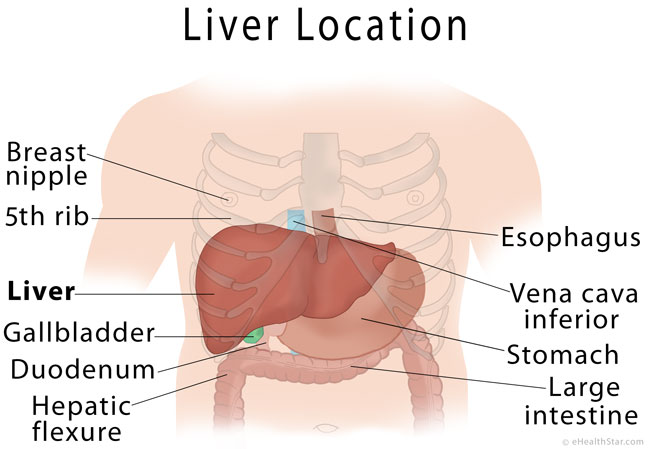
Picture 1. Liver location
Related terms:
- Hepar (in ancient Greek) = liver
- Hepatic = related to the liver, for example, hepatic artery or vein
Gross Liver Anatomy

Picture 2. Liver anatomy diagram: anterior and inferior surface
Liver Size (in Adults)
- The longest vertical length (on the right side) is 6-8 inches (15-17 cm) [6].
- The horizontal length is 8-9 inches (20-23 cm) [6].
- The liver weighs about 1,500 grams (~3 pounds) in average [1,14].
Liver Shape and Parts
The liver has a shape of a triangular pyramid with the base pointing to the side of the lower part of the right rib cage and the apex pointing to the left breast nipple [1,9]. The liver is encapsulated in a fibrous connective tissue capsule (Glisson’s capsule) and almost completely covered by the abdominal membrane (peritoneum), except in the “bare area” on its bottom [6].
The liver consists of 2 main lobes–the right and left–, each of which is divided into 8 segments [1]. The two main lobes are separated by the falciform ligament, which attaches the liver to the abdominal wall and diaphragm [24]. Two smaller lobes–the quadrate and caudate lobe–are on the bottom side of the liver [1].
The liver has 2 surfaces [1]:
- The upper diaphragmatic surface, which is dome-shaped and can be divided into four continuous surfaces: the upper (superior), front (anterior), back (posterior) and right surface
- The bottom, inferior or visceral surface
The liver has a sharp anterior (inferior) border that separates the anterior and inferior surface; other liver borders are rounded [6].
Hilum or porta hepatis is the central part of the liver through which the common hepatic duct (bile duct), hepatic artery and portal vein enter the liver.
Blood Vessels
The liver receives blood from the hepatic artery (20-40%) and portal vein (60-80%) [1,4]. The hepatic artery and portal vein each deliver about 50% of oxygen to the liver [3]. The blood is drained from the liver by the hepatic vein.
- Hepatic artery branches from the celiac artery and this from the abdominal aorta.
- Portal vein collects blood from the stomach, pancreas, gallbladder, small and large intestine and spleen and delivers it to the liver.
- Hepatic vein leads to the inferior vena cava which drains blood to the heart.
- Reference: [6]
Nerves
- Sympathetic nerves arise from the celiac plexus [3,4].
- Parasympathetic nerves arise from the vagus nerve [3,4].
Bile Ducts
The bile ducts form a branched structure called the biliary tree [6].
The bile produced by the liver cells is drained by the bile canaliculi, which join together and eventually form the right and left hepatic duct, which join into the common hepatic duct, which leaves the liver. Common hepatic duct, which joins with the cystic duct from the gallbladder, join together and form the common bile duct, which opens into the duodenum.

Picture 3. Biliary tree
Nearby Organs
The entire upper (diaphragmatic) surface of the liver is covered by the diaphragm. Above the diaphragm, in the chest cavity (thorax), there is the right lung with the corresponding part of lung membrane (pleura) and the heart within the heart sac (pericardium).
The inferior (visceral) surface of the liver is in touch with the gallbladder, right kidney and adrenal gland, stomach, duodenum and “hepatic flexure”–the area where the right vertical (ascending) colon turns into the horizontal (transverse) colon [1,4,9].
The back liver surface touches the esophagus and the inferior vena cava.
Video 1. Liver anatomy with nearby organs
(the main content is between 3.30 and 11.10).
Microscopic Anatomy
Hepatic Lobules
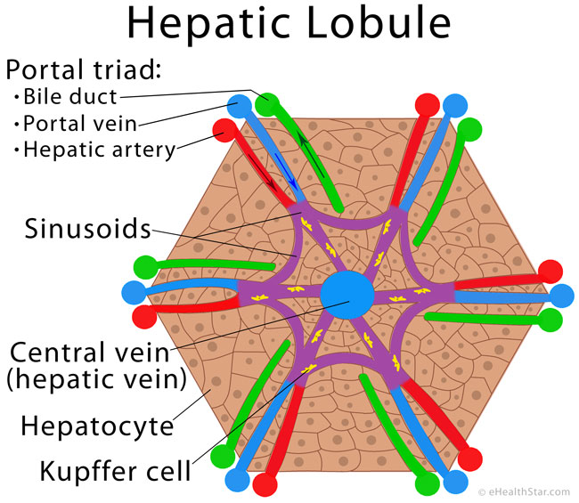
Picture 4. Hepatic lobule
The liver tissue consists of small lobules 1-2.5 mm in size [6]. Each lobule has the central vein (a branch of the hepatic vein), which is surrounded by 6 pairs of vessels consisting of a branch of the portal vein and a branch of the hepatic artery. The blood flows from the hepatic artery and portal vein through the capillaries called the sinusoids into central veins. Accompanying the pair of blood vessels, there are small bile ducts that collect the bile from hepatic cells and carry it out of the liver [15].
Liver Cells
The two most important types of cells in the liver are [2]:
- Hepatocytes are the main liver cells that perform most of the liver’s functions.
- Kupffer cells are a type of cells called macrophages or phagocytes; they remove worn blood cells and microbes from the circulation.
What does the liver do?
The liver produces some substances you need, breaks down harmful substances, drugs and alcohol, removes old red blood cells and some microbes, helps to digest fats, and stores glucose and some minerals and vitamins.
1. Metabolism
a) Glucose metabolism
Excessive glucose that enters the blood after meals is rapidly taken by the liver and stored as glycogen (glycogenogenesis) [26]. Up to 100 grams of glycogen can be stored in the liver [7]. Between meals, when the blood glucose levels fall, glycogen is converted back to glucose (glycogenolysis) and released into the blood. The liver can also produce glucose from non-carbohydrate sources, such as amino acids, lactate, pyruvate and glycerol (gluconeogenesis) [11].
b) Lipid metabolism
The liver breaks down the excessive amount of fatty acids that enter the liver into acetate (beta-oxidation of fats), which can be used for the synthesis of other substances [26]. The liver can also convert the excessive amounts of glucose and amino acids into fatty acids and triglycerides, which are released into the blood and stored elsewhere in the body [26].
The liver produces about 10% of cholesterol that body needs [2,8]. The liver can incorporate cholesterol into lipoproteins and transport it to the tissues. Cholesterol is a precursor of bile acids, vitamin D or steroid hormones: cortisol, aldosterone and sex hormones: testosterone, estrogen and progesterone [28].
The liver handles lipoproteins, such as LDL and HDL cholesterol, which transport fats and cholesterol from and into the liver [24].
Ketones are produced in the liver from fatty acids as an alternative source of energy when there is not enough glucose available for the body cells (in low-carbohydrate diet, starvation or diabetes mellitus).
c) Protein metabolism
The liver can produce nonessential amino acids [26].
During conversion of amino acids into glucose or fatty acids, the liver removes amino groups from amino acids and convert them into ammonia (deamination) and further into urea (urea cycle), which is excreted from the body in the urine [10]. In severe liver disease, ammonia, which cannot be efficiently converted to urea, starts to accumulate in the blood and causes hyperammonemia, which can be life-threatening.
2. Bile Production
The liver produces bile, which is temporarily stored in the gallbladder and then–via the bile ducts–delivered to the first part of the small intestine (duodenum). Bile emulsifies fats and thus enables their digestion and absorption as well as absorption of cholesterol and fat-soluble vitamins A, D, E and K [27].
A severe liver disease, such as hepatitis or cirrhosis, can affect bile production or its delivery to the intestine (cholestasis), which can result in vitamin A, D, E or K deficiency and fat malabsorption with greasy stools (steatorrhea).
The liver produces bilirubin from hemoglobin from worn red cells. Large intestinal bacteria convert bilirubin into stercobilin, which gives a brown color to the stool [2]. In liver diseases with impaired bile production, stool lacks stercobilin, so it has a whitish color.
3. Production of Specific Proteins
The liver produces different proteins listed below.
Prothrombin and fibrinogen are necessary for blood clotting. In severe liver disease, the lack of these proteins increases the risk of bleeding.
Albumins are proteins that help to keep water in the blood. In chronic liver disease, such as cirrhosis, the albumin production can be low, so some water may escape from the blood vessels and start to accumulate in the abdominal cavity (ascites). Albumins also enable transport of minerals (calcium, potassium), fatty acids, bilirubin, hormones (thyroxine) and drugs via the blood.
Globulins carry certain minerals (iron, copper) and hormones via the blood, assist in immune function, etc. [25].
Liver enzymes, such as alanine transaminase (ALT), aspartate transaminase (AST) and alkaline phosphatase, enable synthesis and breakdown of various substances in the liver. Increased or decreased blood levels of liver enzymes can help recognize certain liver diseases.
Reference: [24]
4. Storage of Vitamins and Minerals
The liver stores the following vitamins and minerals:
- Vitamins A [18], D [23], E [19], K [21] and B12 [22] (the stores suffice for several months or years)
- Iron in the form of ferritin and hemosiderin [17]
- Copper [20]
5. Detoxification
The liver removes some harmful substances from the blood and excretes them via the bile [2]. It gradually breaks down some medications, which is why you need to take them repeatedly in order to work. It also breaks down alcohol, which results in a gradual drop of blood alcohol concentration after stopping drinking.
6. Immunity
Kupffer cells, which are a part of macrophage system–along with the spleen and lymph nodes–, help to remove worn blood cells, bacteria, fungi and parasites from the blood [2].
7. Other Functions
- The liver is an endocrine gland; it produces hormones angiotensinogen, thrombopoietin and insulin-like growth factor 1 (IGF-1) [16].
- The liver regularly breaks down insulin and other hormones produced in your body and thus maintains their appropriate concentrations in the blood [2].
Liver Regeneration
The liver has a great regeneration capability; loss or damage of up to 75% of the liver tissue can be completely replaced by healthy liver tissue [5].
Liver Transplantation
You cannot live without the liver. The liver that is irreversibly damaged, for example, due to cirrhosis, can be replaced by the liver from a compatible donor.
Physical Examination
A doctor can evaluate the liver size by percussion and palpation. Percussion over the enlarged liver gives a dull sound.
The normal liver usually cannot be palpated because it is covered by the rib cage. The anterior (inferior) border of the normal liver can sometimes be palpated in thin individuals during deep inhalation [14].
Enlarged liver (hepatomegaly) can be palpated as an abdominal mass below the right rib cage.
- The enlarged, soft and nontender liver without tenderness can be found in fatty liver [13,14].
- The enlarged and tender liver can be observed in liver inflammation (hepatitis), congestive heart failure and cancer [14,12].
- The irregular liver surface can be palpated in cirrhosis and liver cancer [12,13,14].
- The rock-hard liver can be palpated in liver cancer [14].
- References
- Kapoor VK, Liver anatomy Emedicine
- Taylor T, InnerBody
- Luijkx T et al, Liver Radiopedia.org
- Abdel-Misih SRZ et al, 2010, Liver anatomy PubMed Central
- Häussinger D, Liver Regeneration
- Henry Gray, The liver, Anatomy of the Human Body Bartleby
- Wasserman DH, 2008, Four grams of glucose PubMed Central
- King MW, Introduction to cholesterol metabolism The Medical Biochemistry Page
- Henry Gray, Surface markings of the abdomen, Anatomy of the Human Body Bartleby
- Excretion and the liver The Royal Society of Chemistry
- King MW, Gluconeogenesis: endogenous glucose synthesis The Medical Biochemistry Page
- Examination of the liver Stanford University
- Herrine SK, Evaluation of the Patient With a Liver Disorder Merck Manuals
- Wolf DC, Evaluation of the Size, Shape, and Consistency of the Liver National Center of Biotechnology Information
- Introduction to liver SIU School of Medicine
- An Overview Of Endocrine Hormones Secreted By The Liver HGH.org
- Iron storage University of Utah Health Care
- Vitamin A Food and Agriculture Organization of the United Nations
- Vitamin E Georgia Highlands College
- Roberts EA et al, 2008, Liver as a key organ in the supply, storage, and excretion of copper PubMed
- Vitamin K Linus Pauling Institute
- Digestion, absorption and transport of vitamin B12 VeganHealth.org
- Fat-Soluble Vitamins: A, D, E, and K Colorado State University Extension
- Anatomy and Physiology of the liver RnCeus.com
- Globulins Patient.info
- Metabolic Functions of the Liver Colorado State University
- Digestion and absorption of fats University of Washington
- Important Derivatives of Cholesterol Include Bile Salts and Steroid Hormones National Center of Biotechnology Information
- Ronald A et al, Liver AnatomyAtlases
Very helpful information, really enjoyed learning more about the liver