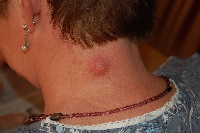
What is epidermoid cyst?
An epidermoid cyst is a benign (noncancerous) encapsulated lesion that arises from the epidermal cells on the skin surface or from the uppermost part of the hair follicle (follicular infundibulum) [3]. Its wall consists of the epidermal cells and its content mainly of the protein keratin and fat [2].
Synonyms for an epidermal cyst include keratin, epithelial, epidermal and follicular infundibular cyst [1,2].
Epidermoid and pilar cysts are more commonly, but technically wrongly, called sebaceous cysts.
Epidermoid Cyst Symptoms
An epidermoid cyst appears as a dome-shaped, firm or fluctuant, fixed lump from few millimeters to 5 centimeters in size, covered by normal, white, yellowish or dark brown skin [2]. It can have a blackhead-like opening, which can ooze a white or yellow greasy and rancid-smelling material [3,4].
Epidermoid cysts most commonly appear on the face, scalp, behind the ear, on the neck, upper back or chest, shoulders, arms, scrotum, vagina and labia [1,2]. Most epidermoid cysts appear in adults [3,6].
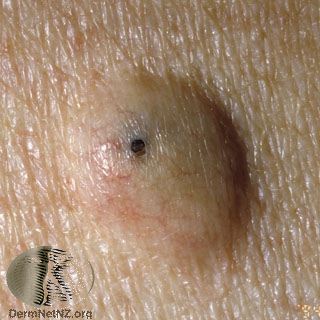
Picture 1. An epidermoid cyst with an opening
(source: DermNet NZ, CC license)
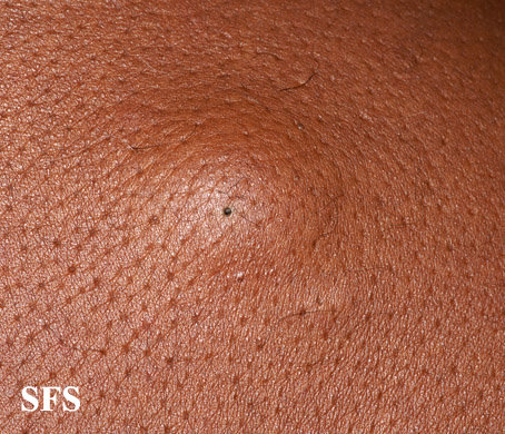
Picture 2. Epidermoid cyst
(source: SF da Silva, MD, Dermatology Atlas)
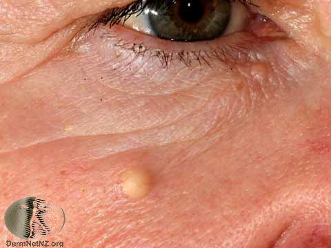
Picture 3. An epidermoid cyst on the face
(source: DermNet NZ, CC license)
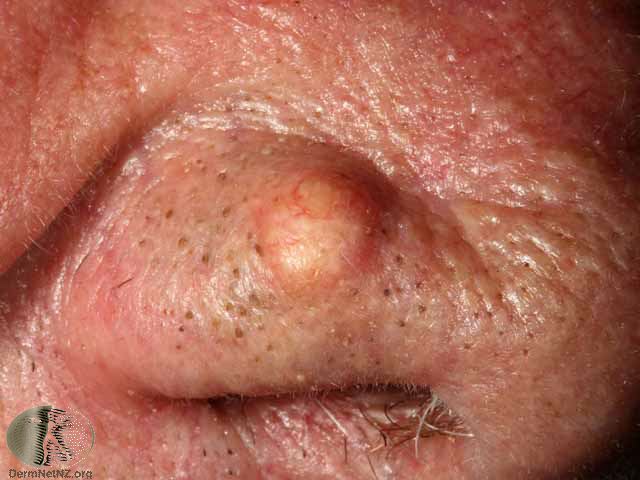
Picture 4. An epidermoid cyst on the eyelid
(source: DermNet NZ, CC license)
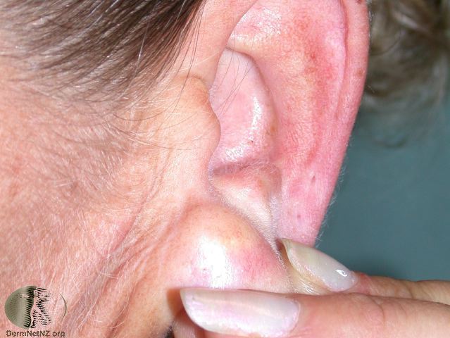
Picture 5. An epidermoid cyst on the earlobe
(source: DermNet NZ, CC license)
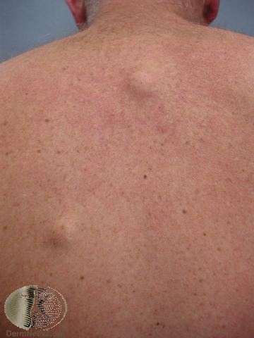
Picture 6. Two epidermoid cysts on the back
(source: DermNet NZ, CC license)
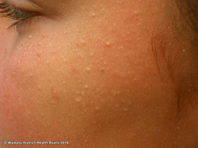 Milia are small (1-2 mm) white epidermoid cysts on the face, mainly in newborns, children and adolescents [3].
Picture 7. Milia on the face of a child
(source: DermNet NZ, CC license)
Epidermal inclusion cyst is a type of epidermoid cyst, which develops from the cells of the surface skin layer (epidermis), when they are pushed into the deeper skin layer (dermis) during trauma or surgery, for example.
Picture 8. An inflamed epidermal inclusion cyst at the back of the neck
(source: Steven Fruitsmaak, Wikimedia, CC license)
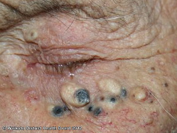 Favre-Racouchot syndrome refers to the development of the solar comedones and epidermoid cysts in the elderly with a condition called elastosis and accumulated sun exposure [3,5].
Picture 9. Solar comedones and epidermoid cysts (Favre-Racouchot syndrome)
(source: DermNet NZ, CC license)
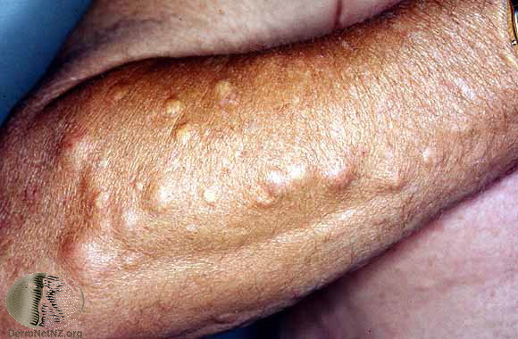 Gardner’s syndrome includes multiple epidermoid cysts, fibromas and lipomas, osteomas and polyps of the colon.
Picture 10. Multiple epidermoid cysts on the forearm as part of Gardner’s syndrome
(source: DermNet NZ, CC license)
Infection
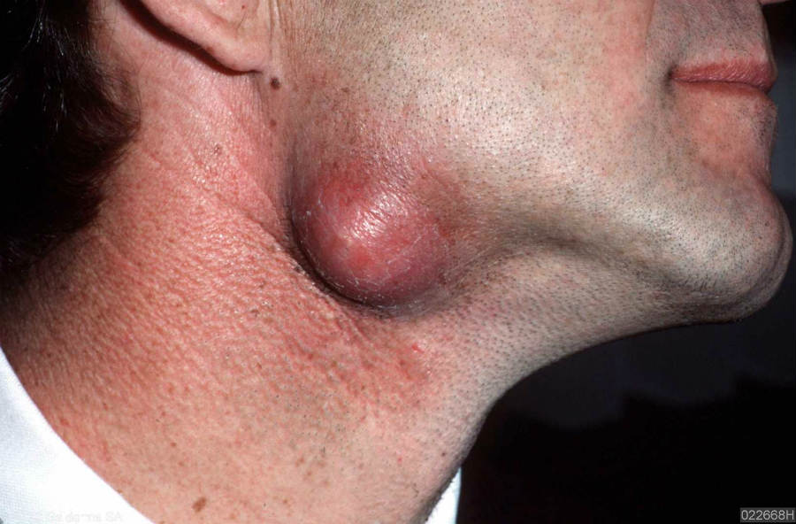 An infected epidermoid cyst is swollen, red, tender and painful.
Picture 11. An infected epidermoid cyst under the jaw
(source: PCDS.org.uk, by permission)
Causes and Risk Factors
The exact cause of epidermoid cysts is not known. Risk factors include acne, trauma, surgery, exposure to UV light or sun, an infection with the Human papillomavirus (HPV) and genetic predisposition (for example, familial adenomatous polyposis) [2,3].
Differential Diagnosis
Other lumps that can look similar to an epidermoid cyst [2]:
- Pilar (trichilemmal) cyst
- Lipoma
- Dermoid cyst
- Ganglion cyst
- Pilonidal cyst
- Enlarged lymph node
- Nodular acne
- Skin cancer (basal or spinous cell carcinoma)
- Steatocystoma
Treatment
An epidermoid cyst can be removed by surgical excision. The removal of both the greasy content and the cyst wall is necessary for the cyst to be cured. There is no known prevention or home treatment for epidermoid cysts. If you only squeeze an epidermoid cyst, it will very likely recur in few weeks or months.
- References
- Epidermal inclusion cyst, clinical presentation Emedicine
- Zuber TJ et al, 2002, Minimal Excision Technique for Epidermoid (Sebaceous) Cysts American Family Physician
- Epidermal inclusion cyst, overview Emedicine
- Epidermoid and Pilar Cysts (Sebaceous Cysts) Patient.info
- Feinstein RP, Favre-Racouchot Syndrome (Nodular Elastosis With Cysts and Comedones) Emedicine
- Oakley A, Cutaneous cysts and pseudocysts DermNetNZ


